昨日Instagramに投稿しました、症例2の解説例(黒字:前田 赤字:内多先生)です。
インスタでの回答結果は
癌:71%、炎症:29%でリンパ腫と回答された方はいませんでした。
Here is an example of the explanation of Case 2 (black letters: Maeda, red letters: Dr. Uchita), which I posted on Instagram yesterday.
The results of the responses on Instagram were.
Cancer: 71%, Inflammation: 29%, no one answered lymphoma.
病変部位をこの写真では言及するのは困難ですが、胃体下部大弯に存在するピロリ除菌後の萎縮粘膜を背景とした7mm程度の領域です。周囲がやや発赤していることで褪色調に見えており、境界は明瞭で形状は不整形です。
It is difficult to mention the site of the lesion in this photograph, but it is an area of about 7 mm with a background of atrophic mucosa after Pylori eradication in the greater curvature of the lower gastric body. The lesion appears faded due to the slightly erythematous surrounding area, and has a clear border and irregular shape.
インジゴカルミン撒布像では白色光観察で視認できた境界と一致してインジゴの溜まりを認めます。しかし、一層外側の少し発赤している領域の外側には境界となる様なインジゴの溜まりを認めません。
The indigocarmine scatter image shows a pool of indigos consistent with the boundary visible in the white light observation. However, outside of the slightly erythematous region on the outer layer, there is no indigo accumulation that would serve as a boundary.
NBI観察でも同様にBrownishな領域として境界が視認できます。
The boundary is also visible as a brownish area in NBI observation.
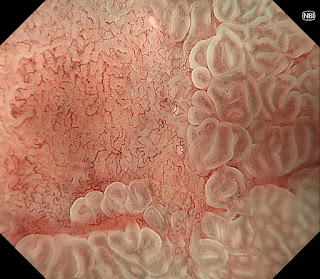
NBI併用拡大観察では背景粘膜にLight Blue Crestを認め腸上皮化生粘膜であることが分かります。また、今までに境界として視認できていた軽度陥凹した箇所に一致してDemarcation Lineを認めます。微小血管構築像は不整でありMV:irregular、表面微細構造は視認できずMS:absentと判断しました。癌と診断しますが未分化型癌を示唆する様な無構造領域も認めず分化型腺癌と考えました。
また、白色光観察にて台状挙上などの所見は認めず深達度Mと診断してESDを行いました。
病理結果は早期胃癌(高分化腺癌:tub1)で深達度はMでした。
The magnified view with NBI shows a Light Blue Crest on the background mucosa, indicating the presence of intestinal epithelialized mucosa. A demarcation line is also observed in line with a slightly depressed area that was previously visible as a border. The microvascular architecture is irregular and is classified as MV: irregular, while the surface microstructure is not visible and is classified as MS: absent. The diagnosis of carcinoma was considered to be differentiated adenocarcinoma, as there were no unstructured areas suggestive of undifferentiated carcinoma.
In addition, no elevation of the pedicle was observed by white-light observation, and ESD was performed with a diagnosis of M depth.
The pathological result was early gastric cancer (highly differentiated adenocarcinoma: tub1) with a depth of M.
毛細血管網を見ていくと軽度の不整形はあるものの、networkを密に形成しており、分化のよい小型の腺管からなる高分化腺癌を考えます。辺縁にはWGAと思われる白色を呈する所見も認めます。NBIではしっかりと境界も認め、腫瘍の診断として難易度は低めです
The capillary network is mildly irregular, but the network is densely formed, suggesting a well-differentiated adenocarcinoma composed of small well-differentiated ducts. NBI shows a well-defined border, making the diagnosis of tumor less difficult.


0 件のコメント:
コメントを投稿