2023年3月4日土曜日

5G遠隔診療の取り組み📲 5G Remote Medical Treatment Initiatives

当院は、神戸大学国際がん医療センター(ICCRC)の森田先生にお誘い頂き、香川大学さんと3拠点4院でNTT docomoの5G回線と映像伝送ソリューション『LiveU』を用いた遠隔診療のプロジェクトに取り組んでいます。

背景として現在我が国はSociety 5.0時代を迎えており、IoT(Internet of Things)により人とモノが繋がり、人工知能(AI)により少子高齢化や地方過疎化等の課題克服を目指した社会の実現が待たれます。

消化器内視鏡分野も発展が著しく、様々な診断・治療技術やそのデバイスが開発される一方で、医師の地域偏在等の理由で受けられる医療や医師教育の質に差が生じているのも事実です。

私たちは前述の取り組みにより、現状打開の一歩にならないかと考えています。


Our hospital, invited by Dr. Morita of Kobe University International Cancer Center (ICCRC), is working with Kagawa University on a project for remote medical care using NTT docomo's 5G line and "LiveU," a video transmission solution, at four hospitals in three locations.

As a background, Japan is currently entering the Society 5.0 era, where people and things are connected through the Internet of Things (IoT), and artificial intelligence (AI) is expected to help overcome issues such as the declining birthrate, aging population, and depopulation in rural areas.

The field of gastrointestinal endoscopy is also making remarkable progress, with the development of various diagnostic and therapeutic technologies and devices, but it is also true that there is a gap in the quality of medical care and physician education available due to the uneven distribution of physicians in different regions.

We hope that the aforementioned efforts will be a step toward overcoming the current situation.


<LiveU送信機>
LiveU Transmitter
バックパックに入るほどのサイズで4K品質の動画伝送が可能です
Small enough to fit in a backpack and capable of 4K quality video transmission


<映像伝送のイメージ>
Image of image transmission
内視鏡術者の内視鏡画面は5G回線を経由してLiveU送信機にてdocomoのクラウドへと伝送されます。それをほぼリアルタイムに各拠点のダブレット端末(iPadなど)やPCなどで受信できます。アノテーション(キャプチャー画像に書き込みなどを行います)を適宜加え、それが発信元も含めた各施設にリアルタイムに共有されるため指導や情報共有が可能となります。
The endoscopist's endoscope screen is transmitted to the docomo cloud via a LiveU transmitter via a 5G line. It can be received almost in real time by a doublet terminal (e.g., iPad) or PC at each site. Annotation (writing on captured images) can be added as needed, and this information is shared in real time with each facility, including the sender, allowing for guidance and information sharing.


↓↓docomoさんが作成して下さった動画です↓↓
Here is a video created by DOCOMO

<内視鏡4K映像伝送実証 1分ver.>
Demonstration of endoscope 4K video transmission 1min. ver.


<内視鏡4K映像伝送実証 3分ver.>
Endoscope 4K image transmission demonstration 3min. ver.
BOSSのインタビューもあります❗️
There is also an interview with BOSS: ❗️


今後も随時Instagram等で進捗状況を発信📲していきます。
We will continue to post progress updates on Instagram and other social media.


2023年3月1日水曜日

症例2📗 Case 2


昨日Instagramに投稿しました、症例2の解説例(黒字:前田 赤字:内多先生)です。
インスタでの回答結果は
癌:71%、炎症:29%でリンパ腫と回答された方はいませんでした。
Here is an example of the explanation of Case 2 (black letters: Maeda, red letters: Dr. Uchita), which I posted on Instagram yesterday.
The results of the responses on Instagram were.
Cancer: 71%, Inflammation: 29%, no one answered lymphoma.

病変部位をこの写真では言及するのは困難ですが、胃体下部大弯に存在するピロリ除菌後の萎縮粘膜を背景とした7mm程度の領域です。周囲がやや発赤していることで褪色調に見えており、境界は明瞭で形状は不整形です。
It is difficult to mention the site of the lesion in this photograph, but it is an area of about 7 mm with a background of atrophic mucosa after Pylori eradication in the greater curvature of the lower gastric body. The lesion appears faded due to the slightly erythematous surrounding area, and has a clear border and irregular shape.

インジゴカルミン撒布像では白色光観察で視認できた境界と一致してインジゴの溜まりを認めます。しかし、一層外側の少し発赤している領域の外側には境界となる様なインジゴの溜まりを認めません。
The indigocarmine scatter image shows a pool of indigos consistent with the boundary visible in the white light observation. However, outside of the slightly erythematous region on the outer layer, there is no indigo accumulation that would serve as a boundary.

NBI観察でも同様にBrownishな領域として境界が視認できます。
The boundary is also visible as a brownish area in NBI observation.

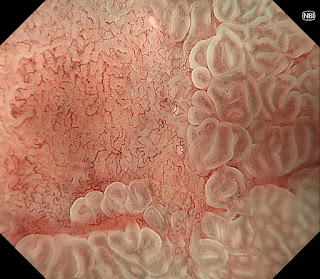
NBI併用拡大観察では背景粘膜にLight Blue Crestを認め腸上皮化生粘膜であることが分かります。また、今までに境界として視認できていた軽度陥凹した箇所に一致してDemarcation Lineを認めます。微小血管構築像は不整でありMV:irregular、表面微細構造は視認できずMS:absentと判断しました。癌と診断しますが未分化型癌を示唆する様な無構造領域も認めず分化型腺癌と考えました。
また、白色光観察にて台状挙上などの所見は認めず深達度Mと診断してESDを行いました。
病理結果は早期胃癌(高分化腺癌:tub1)で深達度はMでした。

The magnified view with NBI shows a Light Blue Crest on the background mucosa, indicating the presence of intestinal epithelialized mucosa. A demarcation line is also observed in line with a slightly depressed area that was previously visible as a border. The microvascular architecture is irregular and is classified as MV: irregular, while the surface microstructure is not visible and is classified as MS: absent. The diagnosis of carcinoma was considered to be differentiated adenocarcinoma, as there were no unstructured areas suggestive of undifferentiated carcinoma.
In addition, no elevation of the pedicle was observed by white-light observation, and ESD was performed with a diagnosis of M depth.
The pathological result was early gastric cancer (highly differentiated adenocarcinoma: tub1) with a depth of M.

毛細血管網を見ていくと軽度の不整形はあるものの、networkを密に形成しており、分化のよい小型の腺管からなる高分化腺癌を考えます。辺縁にはWGAと思われる白色を呈する所見も認めます。NBIではしっかりと境界も認め、腫瘍の診断として難易度は低めです
The capillary network is mildly irregular, but the network is densely formed, suggesting a well-differentiated adenocarcinoma composed of small well-differentiated ducts. NBI shows a well-defined border, making the diagnosis of tumor less difficult.